


Abstract
Purpose: Microbial contamination of manual toothbrushes relative to their design has been documented for decades, citing concern for cross contamination and self-infection with microorganisms. A pilot study of different power toothbrushes was conducted, to compare a solid-head brush to 2 hollow-head brushes for residual contamination with commonly occurring oral microorganisms.
Methods: Participants who met inclusion criteria were enrolled and brushed twice daily for 3 weeks with 1 of 3 randomly assigned power toothbrushes. Brush heads were vortexed and cultured using 5 appropriate media for oral microorganisms: anaerobes and facultative microorganisms, yeast and mold, oral streptococci and oral enterococci anaerobes, Porphyromonas gingivalis, and Fusobacterium species. Analysis of covariance was used to compare the brush groups for transformed microbial counts after adjusting for any demographic variables that may have confounded the results.
Results: The solid-head power toothbrush was found to have significantly less microbial contamination than either of the 2 hollow-head power toothbrushes for all the bacteria tested and less than 1 of the hollow-head brushes for yeast and mold.
Conclusion: The solid-head power toothbrush studied had significantly less residual microbial contamination than the 2 hollow-head power toothbrushes after 3 weeks of twice daily brushing with non-antimicrobial toothpaste.
Introduction
Studies have reported that toothbrushes become contaminated with microorganisms during use, and the amount of these organisms increases with repeated use.1-4 The microorganisms which survive on toothbrushes can be transmitted back to the user during subsequent brushings with the potential for causing further infections.1,2,5,6 In one study, 70% of toothbrushes were found to be heavily contaminated with pathogenic microorganisms after use.7 Most microbial contamination was reported to be within the tufts of bristles/filaments of the multitufted toothbrushes tested. Bacterial survival was dependent upon the type of bacteria (aerobic versus anaerobic) as well as the toothbrush design and bristle/filament type.8,9 Multi-tufted toothbrushes that had the anti-microbial ingredient, Triclosan, added to the heads were not shown to reduce residual contamination, but use of a dentifrice containing Triclosan did reduce it significantly.10-12 Mehta et al found that retention of moisture and oral debris in the bristles, as well as the use of a cap on the brush, increased microbe survival and retention.7
While no studies to date have demonstrated that bacterial growth on toothbrushes can lead to systemic health effects, several microorganisms have been associated with systemic diseases.4,5 For instance, Fusobacterium nucleatum frequently serves as a “bridge bacterium,” promoting plaque formation with other oral pathogens, especially between early and late colonizing bacteria in the oral cavity.13 It has been found in colorectal tumor samples and is being studied for its role in carcinoma, inflammatory bowel disease and early-stage adenomatous polyp lesions, precursors of colorectal cancer.14 Studies have also looked at this organism's role in pre-term birth and stillbirth since it has been found in the amniotic fluids of pregnant women who have miscarried.15,16 Candida species can cause mild to severe infections of the mouth, throat, esophagus and even the brain. In immunocompromised individuals, this infection can even be fatal.17 Streptococcus sanguis and Porphyromonas gingivalis have been shown to induce platelet aggregation, which leads to thrombus formation and the potential for a heart attack or stroke due to an embolus.18 Quantification of oral bacteria has been demonstrated through the use of rapid adenosine triphosphate (ATP) driven bioluminenscence.19
No studies were found to-date reporting on residual microbial contamination of various types of power toothbrushes with different head designs. The purpose of this in-vitro study was to compare the residual microbial contamination of a power toothbrush designed with a solid head with 2 power toothbrushes designed with hollow heads.
Methods and Materials
Approval to conduct this study was obtained from the Committee for the Protection of Human Subjects at the University of Texas Health Science Center at Houston. A convenience sample of 40 subject volunteers were recruited from the dental clinics at the University of Texas School of Dentistry at Houston, were enrolled in the pilot study and agreed to participate by signing the consent form. All participants met inclusion criteria and were stratified by ATP scores and systematically assigned to brush with 1 of the 3 test power toothbrushes shown in Figure 1: 2 hollow-head toothbrushes, Sonicare® DiamondClean (H1) (Philips Electronics, Andover, MA) and Oral-B® ProfessionalCare Smart Series 5000 (H2) (Procter and Gamble, Cincinnati, Ohio) or the solid-head toothbrush, Broxo® Orabrush™ (S) (Advance Response Corp, NY). Due to limits in laboratory resources, the brushes of the first 10 subjects assigned to each group, representing an equal distribution of brush types, were ultimately cultured and included in the final analysis resulting in a sample size of 30.
Inclusion Criteria
Had received an oral prophylaxis within the previous 12 months, but not within the last 4 months
Had not taken a systemic antimicrobial compound for the past 6 months
Had not used a prescription antibacterial mouth rinse in the last 6 months and agreed to abstain from using any mouth rinses during the study
Agreed to brush twice daily with 1 designated toothpaste
Had an ATP driven bioluminescence meter score in the range of 800 to 1000 (CariScreen® Oral BioTech, Albany, Ore)
Was between the ages of 25 to 70 years
Had a minimum of 6 teeth in each quadrant
Protocol
Participants agreed to brush for 2 minutes, twice a day, for a period of 3 weeks. All subjects were instructed to use the assigned toothbrush with designated toothpaste (Crest Cavity Protection®, Procter and Gamble, Cincinnati, Ohio) and to refrain from using any other dental products, such as toothpastes or mouth rinses, for the study duration. No further oral hygiene instructions were offered. Participants were also advised to contact the primary investigator in the event they were prescribed an antibiotic for a medical condition as this would eliminate their results from the study. One participant was withdrawn due the use of an antibiotic nasal spray. All participants were allowed to keep the power brush at the end of the study after submitting the 1 used toothbrush head for testing.
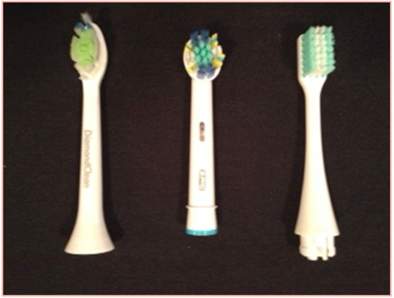
Power Toothbrush Heads
Sonicare® DiamondClean, Oral-B® ProfessionalCare Smart Series 5000 and Broxo®Orabrush™
Toothbrush heads were placed in numbered sterile tubes by the participants at the conclusion of 3 weeks and transported to the lab for testing. Participants from each brush group were tested independently at the beginning and end of the 3 week brushing period using a sterile swab and an ATP driven bioluminescence meter (CariScreen®, Oral BioTech, Albany, Ore) for the purpose of balancing groups for oral hygiene levels. Intact toothbrush heads from each of the 3 brush groups were cultured independently after collection at set times to avoid cross contamination by a research technician blind to study design. The tubes containing the brush heads were allowed to air-dry (250C) for 4 hours prior to processing to simulate regular home use. Ten ml of sterile peptone-saline buffer solution (1% peptone, 7.5% saline, pH 7.0) were added to each of these tubes which were thoroughly agitated for 2 minutes at high speed using a Vortex mixer (Troemner-Henery, Thorofare, NJ). Serial 10-fold dilutions were made in PBS and specimens were plated and incubated in Brewer jars at appropriate temperatures and atmospheres (AnaeroGen Saachets: Fishe-Thermo Scientific that lower oxygen content to no less than 1% within 30 minutes and add 7 to 9% CO2) on 5 different solid microbiological medium:
Brain Heart Infusion agar (Difco, Becton Dickinson and Co., Sparks, MD), a general, non-selective microbial medium for anaerobes and facultative microorganisms (A/FA);
Yeast Mold agar (Difco, Becton Dickinson and Co., Sparks, MD), a selective medium for yeast and mold (Y/M)
Mitis-Salivarius agar (Difco, Becton Dickinson and Co., Sparks, MD), a selective medium for oral anaerobic streptococci and oral enterococci anaerobes (S/EC)
PGING AS 6422 (Anaerobe systems, INC, Morgan Hill, Cali) a selective medium for Porphyromonas gingivalis (PGING)
FSA AS 6427 (Anaerobe Systems, Inc, Morgan Hill, Cali), a selective medium for anaerobic Fusobacterium species. (Fuso)
Because a general count was desired, a selective media was chosen for fastidious anaerobes. Media were checked for quality control with the designated microorganisms. If deemed necessary, 0.1 ml was plated directly from the specimen tubes for PGING and aerobes. Petri plates were appropriately incubated until colonies were large enough to be easily counted. All bacterial media were incubated at 350C and the yeast/mold media at 300C. Plates containing 100 to 300 colony forming units (CFU) were selected for counting. Values of 0 for the microbial counts were converted to 1 prior to log10 transformation. This resulted in transformed values of 0 for those with no recoverable colonies. On occasion, Gram-stained slides of organisms from colonies were observed. After use, all experimental materials were disposed of according to the University Infection Control policy.
Statistical Analysis. Prior to statistical analysis, total microbial counts (in the 10 ml specimen) were converted to log10 to approximate a normal distribution for the data. Descriptive statistics in the form of means and standard deviations were calculated for the transformed data. Comparisons of the 3 brush groups for demographic characteristics were conducted with one-way analysis of variance (ANOVA) for continuous variables and Fisher's exact test for categorical variables. Analysis of covariance was used to compare the brush groups for transformed microbial counts after adjusting for any demographic variables that may confound the results.
Results
Comparisons of the 3 groups by age and baseline ATP measure using ANOVA, as shown in Table I, indicated that there were no significant differences between the groups with regard to age and baseline ATP measure (p=0.78 and 0.74, respectively). Group comparisons by gender and race using Fisher's Exact Test found there was no significant difference by gender (p=0.66), but there was a significant difference between groups by race (p=0.045). Because of this, further between groups comparisons included race as an independent variable to account for its possible effect as a confounder.
Table II shows group means and standard deviations (in log10) for the microorganisms studied in the 3 brush groups (10 brushes each). Mean microbial counts were lower in the S group than in the H1 or H2 groups in 9 out of 10 comparisons. Microbial levels were higher in the H2 group than in the H1 group in 4 out of 5 comparisons.
Table III shows results of a statistical comparison of the 3 brush head groups for each of the 5 microbial groups. Counts in the S group were significantly lower than in the 2 H groups in 8 out of 10 comparisons (p<0.05). The mean value for the Y/M microbe group was significantly lower for the H1 brush group than in the H2 and S groups. The findings are as follows:
A/FA: Group S significantly lower than H1(13x) and H2 (115x)
S/EC: Group S was significantly lower than H1 (48x) and H2(138x)
Fuso: Group S was significantly lower than H1 (3162x) and H2 (550x)
PGING: Group S was significantly lower than H2 (50x)
YM: all 3 groups were significantly different from each other, with H1 the lowest, S in the middle, and H2 the highest
Data are reported as the total number of microorganisms found in the initial 10 ml tube containing the toothbrush head after vortexing. Statistical significance was set at p<0.05 for the intergroup comparisons.
Discussion
The results of the study indicate that the solid-head power brush had fewer residual microorganisms in general than the brushes with hollow heads. Perhaps the hollow heads provided more surface area for the microorganisms to form biofilms. Less microbial growth on the solid-head power toothbrush could offer a simple solution to the residual microbial contamination problem cited in previous studies.7-9 This information could be especially important for immunosuppressed patients who are extremely vulnerable to pathogenic microorganisms such as Fusobacterium nucleatum that have been shown to contaminate toothbrushes.13
Demographic Group Comparisons For Mean Age, ATP, Sex And Race
Group Means and Standard Deviations (Log10) For Microbial Counts
Additionally, even though the results did not indicate a statistically significant difference between H1 and S with regard to Porphyromonas gingivalis levels (p=0.051, reflected in the 95% confidence interval, −0.004, 3.47, where 0 is contained within the interval), the data indicate a “borderline significance” with a trend for S to be lower than H1 for Porphyromonas gingivalis.
The limitations of this study include the fact that the convenience sample size was small, participant compliance may have been an issue and toothbrush head design factors could have had effects on the outcomes as stated in previous studies.8,9 Participants were advised and given written instructions to avoid the use of other dental products and to brush twice a day, but as with most clinical studies, compliance could only be monitored through self-reports. The toothbrush head design factors which could have contributed to the results may have included the overall number of filaments and number per tuft, the filament construction and material, the size of the head, the storage of the toothbrush and the use of a cap on the brush after brushing. Another factor may have been the total plaque actually removed by each type of toothbrush, but no plaque assessment was performed to affirm or negate. Additionally, had one brush removed more bacteria on a daily basis, there would have been fewer bacteria in the mouth to contaminate the brush. Future studies could control for these variables when comparing residual microbial contamination of solid-head manual with solid-head power toothbrushes and compare the reduction in levels of intra-oral microorganisms as well.
Statistical Comparison of Microbial Counts For the 3 Toothbrush Heads In Each of the 5 Microbial Groups
Conclusion
The solid-head power toothbrush studied had significantly less residual microbial contamination than the 2 hollow-head power toothbrushes after 3 weeks of bi-daily brushing with non-antimicrobial toothpaste.
Footnotes
-
Donna W. Morris, RDH, MEd, is a Professor in the Department of Periodontology and Dental Hygiene at the University of Texas Health Science Center School of Dentistry at Houston, Texas. Millicent Goldschmidt, MS, PhD, is a Professor Emerita in the Department of Diagnostic and Biomedical Sciences at the University of Texas Health Science Center School of Dentistry at Houston, Texas. Harris Keene, DDS, is a retired Professor from the Department of Head and Neck Surgery at the M.D. Anderson Cancer Center at the University Of Texas Health Science Center at Houston, Texas. Stanley G. Cron, MSPH, is a Research Instructor, Center for Nursing Research, School of Nursing, the University of Texas Health Science Center at Houston.
-
This study supports the NDHRA priority area, Clinical Dental Hygiene Care: Assess the use of evidence-based treatment recommendations in dental hygiene practice.
-
Disclosure
This study was funded in part by a grant from Advance Response Corp., NY, NY who provided both products and funding for laboratory supplies. The authors acknowledge that there are no conflicts of interest to report associated with this study.
- Copyright © 2014 The American Dental Hygienists’ Association


