


Abstract
Purpose The purpose of this article is to present a case report of a periapical lesion found in a 24-year-old female who presented to a university dental hygiene clinic with a chief complaint of vestibular pain around tooth #22. Radiographically, the lesion appeared radiolucent and unilocular with well-defined borders and significant bone resorption. The area of radiolucency increased in size over a period of five weeks indicating an aggressive pathosis. Given the clinical and radiographic presentations, the differential diagnosis included an odontogenic keratocystic tumor (odontogenic keratocyst), central giant cell granuloma, periapical cyst, and squamous cell carcinoma. Biopsy revealed the diagnosis of a focal abscess. The patient’s presenting signs and symptoms are reviewed. Management and contributing factors are discussed.
INTRODUCTION
When infection occurs at the apices of teeth, activating the inflammatory response, polymorphonuclear neutrophils and other leukocytes attempt to destroy the pathogens.1 However if the initial inflammatory response fails to clear the pathogens, further tissue degranulation, necrosis, and an increased neutrophil response may result in the formation of a periapical abscess.1 Dental caries is the most common contributor to periapical abscess formation, followed by trauma and root canal treatment failure.2 Left untreated, these lesions can cause extensive bone loss; track into the neck space in the mandible; or ascend to the intracranial sinuses in the maxilla, thus posing significant risks to patients.3,4 Additionally, emergency room visits for dental related complications in the United States were reported at two billion dollars in 2017 and emergency room care for dental infections remains an ongoing problem.5 Over 85% of the dental related complications reported from emergency room care were due to diseases of the pulp and periapical tissues (i.e. dental abscesses).5
Increased prevalence of periapical abscesses has been documented in persons with chronic conditions such as systemic lupus erythematosus, hypertension, rheumatoid arthritis, and diabetes mellitus; and more specifically in populations using glucocorticoid and anti-inflammatory immunosuppressive medications.6,7,8,9 In addition, vitamin D deficiency has also been correlated with periapical cyst formation.10 The purpose of this clinical case study was to discuss a periapical lesion found in a 24-year-old female who presented to a northwestern university dental hygiene clinic with the chief complaint of vestibular pain around tooth #22.
CASE DESCRIPTION
A 24-year-old female presented for care to a northwestern university dental hygiene clinic in February 2022 with a chief complaint of pain in the vestibule adjacent to tooth #22. The patient reported that the pain symptoms began about 8 months earlier and that she experienced periods of exacerbation and remission of the symptoms. Notable findings in the health history included heavy tobacco product usage, including vaping, for four years. The patient reported quitting vaping six months prior to the dental hygiene appointment. Also noted were a poor diet, infrequent dental hygiene care intervals, and an anterior crossbite. The patient denied using any medications, including birth control, and there was no reported history of oral trauma. Finances and dental anxiety played a significant role in the delay of dental care; the last reported dental visit was in 2018.
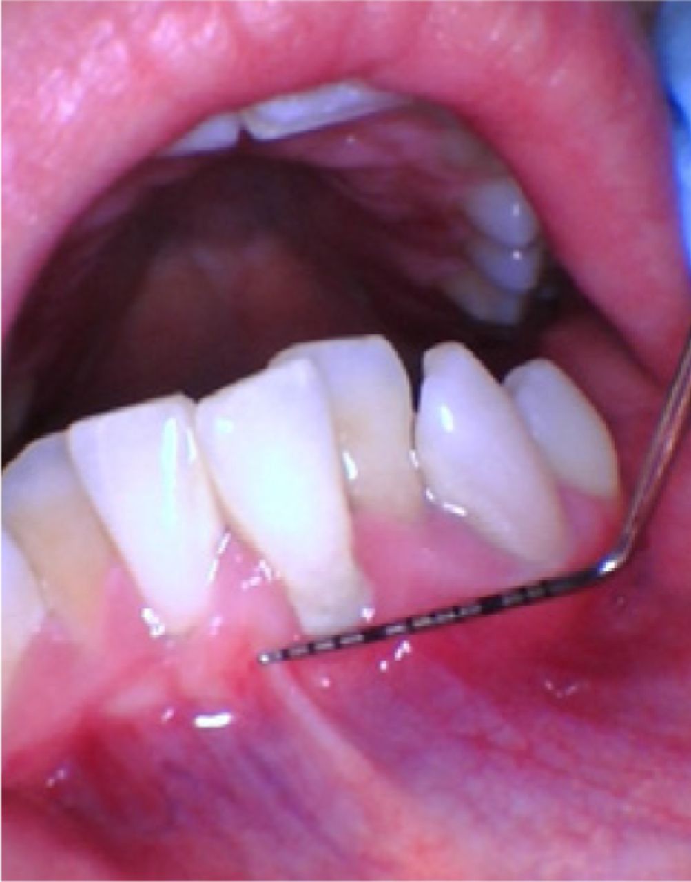
The patient stated that the initial onset of symptoms included deep “throbbing” and “pressurized” pain lasting several days followed by periods of remission and exacerbation, each lasting approximately one to two weeks. Intraoral evaluation revealed a mild, palpable swelling on the floor of the mouth; no color changes were identified. The patient stated the lingual enlargement did not feel abnormal. No significant findings were identified on the extraoral evaluation. The initial periodontal evaluation findings included generalized moderate to severe bone loss, seven sites of bleeding on probing, one site of exudate (#24 buccal), and generalized class 1 mobility of the mandibular anterior teeth (Figure 1). The periodontal diagnosis was generalized stage II, grade B. Teeth #s 22, 24-26 tested vital; tooth #23 tested non-vital.

Intraoral photograph showing exudate on tooth #24 buccal.
Radiographically, the lesion was radiolucent and unilocular with well-defined borders and significant bone resorption. Over the five-week period of dental hygiene care the radiolucency increased in size causing continual bone resorption, indicating aggressive pathosis. Periapical radiographs revealed a large radiolucency encompassing teeth #s 22-24 with vertical bone loss (Figure 2). After comparing the initial periapical radiograph taken on February 28, 2022, with a panoramic image taken March 13, 2022, the radiolucency spanned teeth #s 22-26 (Figure 3). Cone-beam computed tomography (CBCT) was performed and the findings indicated that the lesion appeared to have extended through facial and lingual portions of the mandible, causing complete resorption of the local bone. The CBCT also indicated the radiolucency extended more medially showing a burrowing appearance with significant bone resorption of surrounding teeth # 25 and 26 (Figure 4).

Periapical radiolucency of teeth #s 22-25.

Panographic view of teeth #22-26.

CBCT scan indicating the medial extension of the radiolucency
Differential diagnosis
Given the clinical and radiographic presentations, the differential diagnosis included an odontogenic keratocyst (OKC), central giant cell granuloma (CGCG), and squamous cell carcinoma (SCC). The OKC is also known by a new term that has been proposed for this lesion, keratocystic odontogenic tumor (KCOT). Regardless of the terminology used, the KCOT is a benign developmental tumor or cyst with an aggressive potential. The KCOT/OKC presents as a unilocular or multilocular radiolucency, occurring most often in the posterior mandible molar region.11,12 Most KCOT/OKC tend to occur in the second and third decades. They are discovered during routine radiographs; however, some patients present with pain, swelling and drainage which may represent a secondary infection of the cyst.13 Other lesions may be misdiagnosed as periapical abscesses.14 As the KCOT/OKC lesion progresses, it can cause expansion of the lingual plate and lingual plate perforation. Simple enucleation of the KCOT/OKC results in a high recurrence rate of 25% to 60%.11 Newer treatment approaches include the use of chemical agents or cryotherapy with enucleation to reduce the recurrence rate.11
The central giant cell granuloma accounts for approximately seven percent of benign tumors of the jaw affecting the mandible more frequently than the maxilla. This lesion usually affects individuals under the age of 30 and has a female predilection. The majority of CGCG occur anterior to the first premolar. Early lesions are asymptomatic, but lesions may become expansive over time.15,16 Radiographically, CGCG appears as a radiolucency with sclerotic margins; larger lesions may present with multilocularity, rapid hollowing out of bone, cortical expansion, thinning and perforation, root resorption, and displacement of adjacent structures.15,16 Treatment varies based on clinical characteristics and behavior and may include surgical excision, cryotherapy, enucleation, aggressive curettage with or without chemical cauterization, intralesional steroid injections, calcitonin injections and subcutaneous alpha interferon.17,18 Recurrence rates of 15-20% have been reported by Pogrel while Yadev et al., indicated recurrence rates vary between 13-49%.18,19
In the early stages, squamous cell carcinoma may be asymptomatic, however as this disease progresses pain and mobility may be present.20 Radiographically, SCC presents as a rapidly growing and expansile radiolucent lesion with poorly defined margins.21 Treatment of SCC is based on staging of lesions and may include surgery, radiotherapy and chemotherapy alone or in combination. The prognosis depends on the staging and location.22
Diagnosis and Treatment
Approximately three months after the initial presentation, an oral surgeon performed a biopsy of the areas adjacent to tooth #s 22-24 was performed to rule out any malignancy. Biopsy results indicated the presence of granulation tissue composed of delicate fibrous stroma supporting fibroblasts, small capillaries and moderately dense chronic inflammatory tissue, signifying periapical abscess, and no cancerous pathology.
Due to the significance of oral tissue damage, extensive treatment would be required for the patient to achieve clinically healthy results. Broad-spectrum antibiotics and corticosteroids were prescribed for the treatment of the infection and inflammation. This course of treatment was considered aggressive but was ultimately successful. Restorative treatment planning included additional vitality testing followed by either root canal therapy or extractions of the affected teeth. Four quadrants of scaling and root planing (SRP) were recommended due to probing depths and bleeding sites. The patient did not complete restorative treatment or periodontal therapy as recommended due to dental anxiety and financial considerations. The patient returned for follow-up dental hygiene care in November 2022. A panoramic radiograph was taken (Figure 5). Some bone regeneration was evident, but radiolucency was still present around tooth #s 23 and 24. The clinical examination revealed mild gingival edema but no evidence of exudate or palpable lingual swelling.

Panographic image post antibiotic and corticosteroid treatment.
DISCUSSION
The diversity of possible pathological origins in a case consistent with an odontogenic infection is difficult to identify. Historically, odontogenic infections are comprised of anaerobes including but not limited to: Staphylococcus, Fusobacterium, Prevotella, Bacteroides and Treponema.23 To isolate specific bacterium in this case, a culture would have been taken at the time of biopsy. Due to the initial diagnosis of possible malignancy, bacterial cultures were not ordered.
Fibroblasts found within the biopsy indicated the formation of an extracellular matrix which were responsible for the initial formation of granular tissues. The delicate fibrous stroma supporting fibroblasts, small capillaries and chronic inflammatory tissue indicated stage 3 (proliferative) healing. In the presence of a chronic infection similar to this case, there is a greater likelihood of possible error in the healing cascade, resulting in persistent formation of granulation tissue. Persistent granulation tissue is accompanied by excess fibroblasts and inflammatory cells. Excess reformative cells in combination with inflammatory factors may result in biofilm (infectious toxin) production. Biofilm production generates a perpetual inflammatory response leading to continued granulation tissue formation or extreme delayed healing.24 The patient in this case study experienced periods of exacerbation and remission over the course of six to eight months creating the delayed healing.
The patient’s chronic nicotine use may have contributed to the initial response to infection. Long term histological oral health outcomes related to vaping are still under investigation. Traditional cigarettes with equivalent nicotine levels and carcinogenic effects have been studied and are thought to carry similar potential effects.25 Current literature suggests a link between gingival fibroblasts and delayed healing due to anaerobic bacteria causing damage to the DNA that facilitates wound healing.26 In addition, evidence supports an impaired gas exchange in those who smoke tobacco products leading to systemic hypoxia.27 A hypoxic state is consistent with the presence of anaerobes as previously discussed leading to further periodontal involvement and delayed wound healing.
The combination of socioeconomic factors and a dental phobia contributed to the patient’s problem. The patient was unaware of local programs and resources available to help address financial barriers. A significant back injury sustained during military service required extensive treatment and led to a generalized fear of medical and dental care. These barriers contributed to the disease progression which eventually led to irreversible bone loss.
CONCLUSION
Oral healthcare providers are the frontline defense for identifying lesions that can impact a patient’s oral health, systemic health, and quality of life. In this case study, the patient presented with an aggressive lesion that mimicked a potentially serious condition. Biopsy confirmed an infectious process rather than a malignancy. Delayed healing was present with periods of exacerbation and remission which may have been complicated by a past history of use of tobacco products. Economic constraints, lack of knowledge of financial and community resources, and patient fears of medical and dental procedures contributed to the delayed diagnosis and overall health outcome. Oral health related quality of life is significantly impacted by these burdens and while an immediate oral health condition might be met, other patient needs are not addressed. This case provided an opportunity to learn how multiple factors can impact the progression of an extensive infection.
Footnotes
NDHRA priority area, Client level: Basic science (dental hygiene diagnosis)
- Received October 17, 2022.
- Accepted January 20, 2023.
- Copyright © 2023 The American Dental Hygienists’ Association


